Home » Without Label » Human Body Bones Diagram - Human Skeleton Bones Cut And Paste Activity Teacher Made - This can lead to many health issues such as bone thinning, kidney damage, heart problems and even death.
Human Body Bones Diagram - Human Skeleton Bones Cut And Paste Activity Teacher Made - This can lead to many health issues such as bone thinning, kidney damage, heart problems and even death.
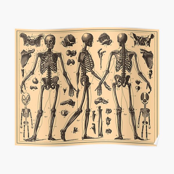
Human Body Bones Diagram - Human Skeleton Bones Cut And Paste Activity Teacher Made - This can lead to many health issues such as bone thinning, kidney damage, heart problems and even death.. Bones in human body provide basic structural shape and support. The bones are a solid structure made up of calcium phosphate and collagen. Herniated disc (slipped disc) transverse foramen. The human skeletal system consists of all of the bones, cartilage, tendons, and ligaments in the body. This framework consists of many individual bones and cartilages.
The bones provide a structural framework and protection to the soft organs. Human body and skeletal system flat illustrations set. This framework consists of many individual bones and cartilages. However, as a child grows, some of the bones fuse together. A forearm bone, it runs from the elbow to the thumb.
Human Anatomy Posters Redbubble from ih1.redbubble.net Human anatomy bones worksheets are a fun and useful way to simply help students understand the anatomy of these body. Herniated disc (slipped disc) transverse foramen. The patella and the pisiform bone of the carpals are the only sesamoid bones that are counted as part of the 206 bones of the body. When autocomplete results are available use up and down arrows to review and enter to select. It is 2 feet long and hollow, to make it lighter. It is composed of 300 bones at birth, but later decreases to 80 bones in the axial skeleton and 126 bones in the appendicular skeleton. Human body, the physical substance of the human organism. The femur or the thigh bone is closest to the body.
12 photos of the human back bone chart.
Also, they are living tissues that grow. This article looks at the anatomy of the back, including bones, muscles. Learn anatomy as you browse our collection of colorful, large and clearly labeled human body diagrams. It is very strong to support the body's weight. Temporal bone occipital bone mandible humerus femur tibia calcaneus fibula ulna radius scapula clavicle scapula. License image the clavicle as viewed from above. The bones are a solid structure made up of calcium phosphate and collagen. Characteristic of the vertebrate form, the human body has an internal skeleton with a backbone, and, as with the mammalian form, it has hair and mammary glands. The long bones of the body contain many distinct regions due to the way in which they develop. Learn more about the composition, form, and physical adaptations of the human body. It is 2 feet long and hollow, to make it lighter. Human heart pilot anatomy 50 x 67 anatomical diagram of bones of the upper body. Anatomy bones diagram, axial skeleton bones, bones of the skeleton quiz, human body bones diagram, labeled diagram skeleton, skeleton diagram with bone names, skeleton system bones, skull bones diagram, human anatomy, anatomy bones diagram, axial skeleton bones, bones of the skeleton quiz, human body.
Human body and skeletal system flat illustrations set. Characteristic of the vertebrate form, the human body has an internal skeleton with a backbone, and, as with the mammalian form, it has hair and mammary glands. There are numerous types and combinations of these worksheets, and they can be found in virtually every medical classroom, no matter size or age the students. Human body, the physical substance of the human organism. On this page, you will find two images i created that illustrate the parts of a long bone and long bone structure.
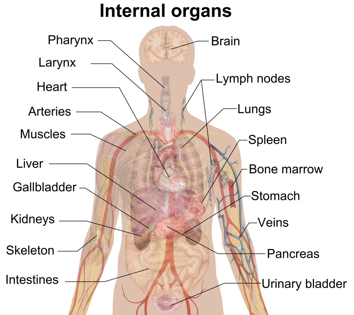
Organ Biology Wikipedia from upload.wikimedia.org A forearm bone, it runs from the elbow to the thumb. The muscle acts as the effort force; Also, they are living tissues that grow. The vertebral column of the lower back includes the five lumbar vertebrae, the sacrum, and the coccyx. The femur or the thigh bone is closest to the body. On this page, you will find two images i created that illustrate the parts of a long bone and long bone structure. In these labeled examples, a human femur is represented without identifying many of the unique characteristics that help differentiate the femur bone from other bones in the human body. Human heart pilot anatomy 50 x 67 anatomical diagram of bones of the upper body.
The ilium is the big bone of the hip, the ischium is the bone on which one sits and the pubis forms the lower frontal hip bone as seen in the diagram.
This diagram depicts picture of female reproductive system diagram 1024×1204 with parts and labels. Craniology is the study of the head and face. On this page, you will find two images i created that illustrate the parts of a long bone and long bone structure. The vertebral column of the lower back includes the five lumbar vertebrae, the sacrum, and the coccyx. This article looks at the anatomy of the back, including bones, muscles. Osteology is the study of the human skeleton, which includes all bones of the body. Posted on june 7, 2016 by admin. The clavicle joins the sternum at the sternoclavicular joint. License image the clavicle as viewed from above. Touch device users, explore by touch or. Joints are points where a muscle is connected to two different bones and contracts to pull them together. 11 photos of the skeleton bones diagram. The bones provide a structural framework and protection to the soft organs.
Posted on june 7, 2016 by admin. The back supports the weight of the body, allowing for flexible movement while protecting vital organs and nerve structures. 12 photos of the human back bone chart. Posted on august 7, 2015 by admin. Learn more about the composition, form, and physical adaptations of the human body.
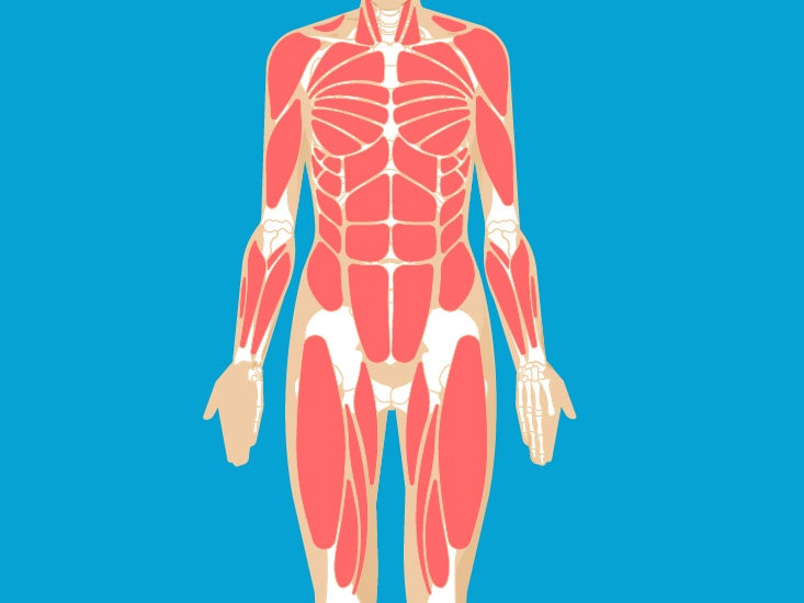
Muscular System Anatomy Diagram Function Healthline from post.greatist.com Learn anatomy as you browse our collection of colorful, large and clearly labeled human body diagrams. This can lead to many health issues such as bone thinning, kidney damage, heart problems and even death. The femur or the thigh bone is closest to the body. Posted on august 7, 2015 by admin. A forearm bone, it runs from the elbow to the thumb. This bone runs down from the shoulder socket and joins the radius and ulna at the elbow. It is very strong to support the body's weight. Herniated disc (slipped disc) transverse foramen.
Posted on january 9, 2021 by kids.
It is composed of 300 bones at birth, but later decreases to 80 bones in the axial skeleton and 126 bones in the appendicular skeleton. This diagram depicts picture of female reproductive system diagram 1024×1204 with parts and labels. It is very strong to support the body's weight. The bones of the pelvis and lower back work together to support the body's weight, anchor the abdominal and hip muscles, and protect the delicate vital organs of the vertebral and abdominopelvic cavities. Temporal bone occipital bone mandible humerus femur tibia calcaneus fibula ulna radius scapula clavicle scapula. Touch device users, explore by touch or. This difference in the number of bones helps forensic anthropologists in. On this page, you will find two images i created that illustrate the parts of a long bone and long bone structure. The number of bones in the human body at birth is 300. 12 photos of the human back bone chart. Posted on august 7, 2015 by admin. The bones provide a structural framework and protection to the soft organs. The longest and the strongest bone in the human skeletal system as you can observe in the labeled skeleton diagram of the human body.